Subjects
Grades
The eye is a complex organ composed of multiple specialized structures. The structures work together to capture light rays and transform them into nerve impulses that the central nervous system can analyze so that we can see.
Among the different types of electromagnetic radiation, only visible light can be perceived by the human eye.
The eye is made up of membranes, transparent structures, and multiple other structures. The eye is also protected by several accessory organs.
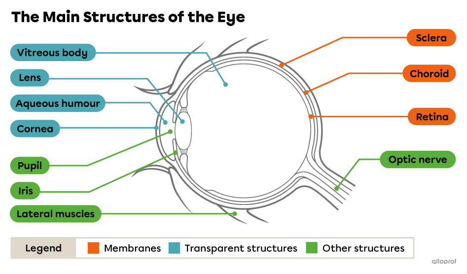
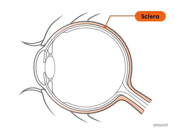
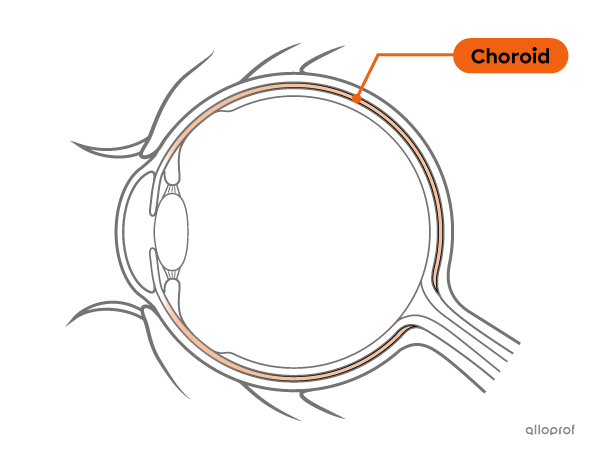
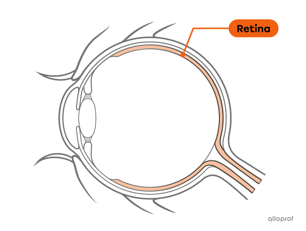
The eye is composed of three superimposed membranes, namely the sclera, the choroid, and the retina.
The sclera, commonly called the white of the eye, is a white membrane that gives shape to the eye.
The choroid is a membrane lining the inside of the sclera. The blood vessels that run through it allow a supply of nutrients and oxygen to the various membranes and structures of the eye.
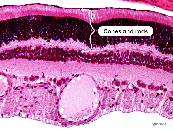
The retina is a membrane lining the choroid and the back of the eye. It includes photoreceptors, cells that capture light and transform it into nerve impulses.
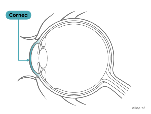
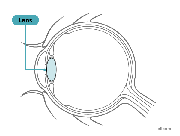
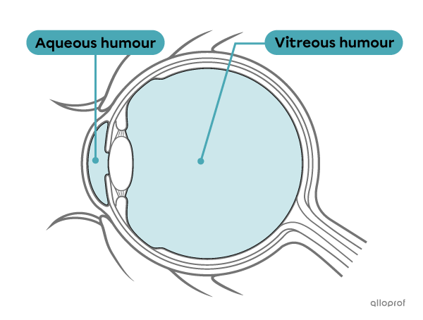
The eye is composed of four transparent structures: the cornea, the aqueous humour, the lens, and the vitreous humour (or vitreous body).
The cornea is a transparent cellular structure located at the front of the eye. The cornea is curved, which means that it acts like a lens: it converges the light rays that enter the eye.
The lens is oval shaped and transparent. Since it has two domed surfaces, it is said to be a biconvex lens. In addition, the lens has the ability to change its shape to adjust the convergence of light rays. It is called lens accommodation.
The aqueous humour and the vitreous humour (or vitreous body) are two transparent liquids. The vitreous humour has a jelly-like consistency and the aqueous humour is more liquid, like water. One of their roles is to allow the eye to maintain its shape by exerting pressure on its various structures. Additionally, both fluids include nutrients and oxygen to nourish the structures of the eye. The aqueous humour and vitreous humour are regularly renewed, which allows the eye to get rid of waste.
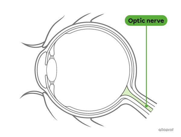
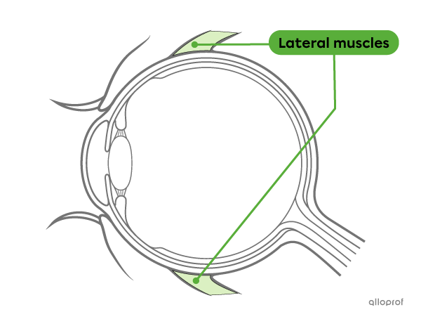
The eye includes many other structures such as the iris, the pupil, the optic nerve, and the lateral muscles.
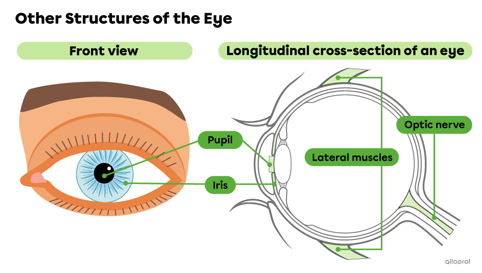
The iris is the coloured structure of the eye. It has a hole called the pupil. Like the diaphragm of a camera, the iris can open or close to adjust the amount of light that enters the eye through the pupil.
The optic nerve is a nerve that connects the eye to the central nervous system. It carries nerve impulses to the brain.
The lateral muscles are muscles surrounding the eyeball. They control the eye movement.
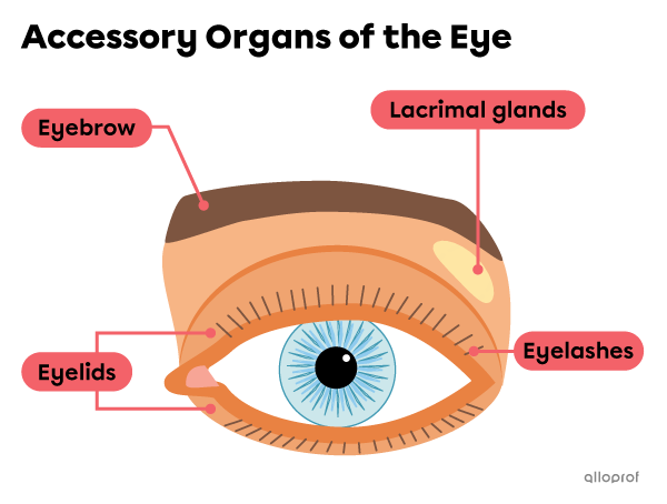
The eye is a complex and fragile system. The accessory organs around the eye provide protection.
The eyelids protect the eyes by covering them. Eyelashes and eyebrows protect the eyes from sweat, dust, and other unwanted elements. In addition, lacrimal glands secrete tears that help clean, lubricate, and moisturize the eye, as well as protect it against bacterial infections. Blinking also helps disperse tears.
Moments in the video:
Objects must emit or reflect visible light to be seen by the human eye. Light rays travel in a straight line to reach our eye.
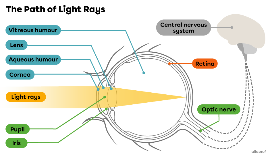
When light rays reach the eye, they first pass through the cornea which causes them to converge. The rays then pass through the aqueous humour and the pupil to reach the lens. The lens also converges light rays. They then pass through the vitreous humour to finally reach the retina.
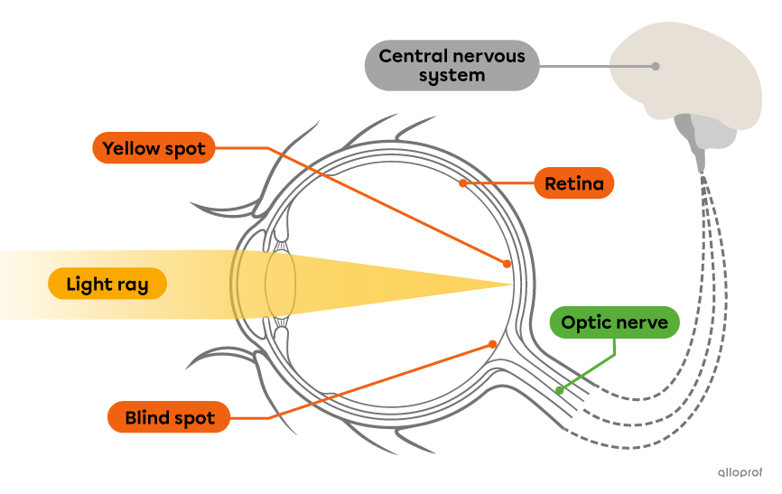
The retina is the light path’s ending point. The cells of the retina actually transform the light rays into nerve impulses which are conveyed by the optic nerve to the central nervous system. The nervous system then analyzes the information received and interprets it in the form of images.
The retina includes multiple cells, called photoreceptors, that capture light and transform it into nerve impulses: the photoreceptors are called the cones and rods.
Structures of the retina and the optic nerve
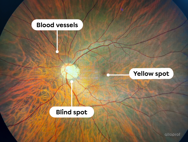
The cones are concentrated in an area called the yellow spot. They capture the colour of images. The rods make it possible to capture the contrast in the images, which is especially helpful in dim environments. They are mostly found around the edges of the yellow spot.
Where the retina attaches to the optic nerve, there are no photoreceptors, so no image is produced if the rays converge on this area. It is why the area is called the blind spot.
Tissues of the retina
Various structures of the retina
For the image perceived by the nervous system to be sharp, the light rays must reach a specific location on the retina. To do this, the lens changes in shape, changing the angle of refraction of the light rays. The lens is said to be accommodating.
So, when an observed object is moved away from the eye, the lens is at rest and has a flattened shape. When the object observed is close to the eye, the lens tends to bulge. Some vision disorders may result in the inability of the lens to sufficiently accommodate in order to create sharp images.
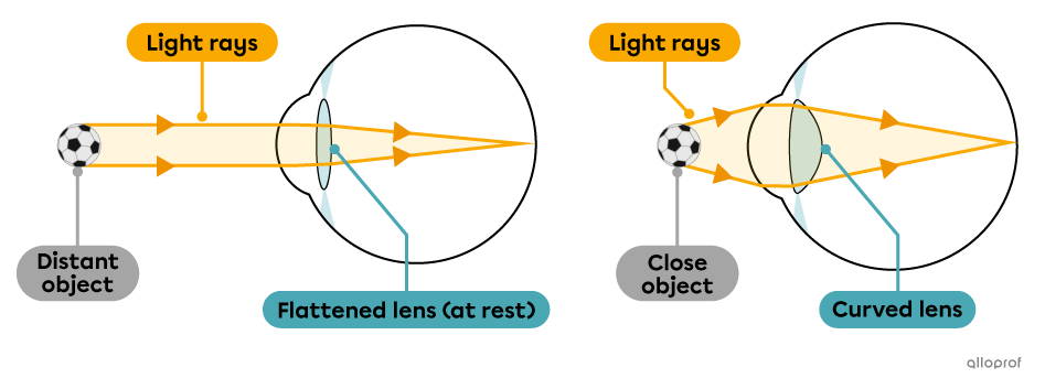
Lens accommodation
In the previous images, the light rays seem to intersect at a single point on the retina. However, the light rays intersect shortly before reaching the retina.
Since the light rays intersect, the image formed on the retina is inverted. The brain compensates and makes us believe the image is right side up.
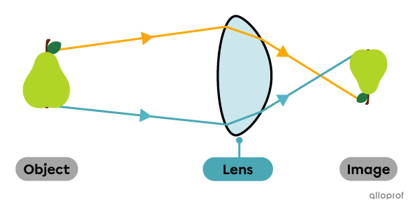
Image inverted by the lens
Moments in the video: