Subjects
Grades
The content covered in this concept sheet goes beyond high school material. It is an enrichment for those who are curious to learn more. For the content covered in Secondary 3, take a look at this concept sheet.
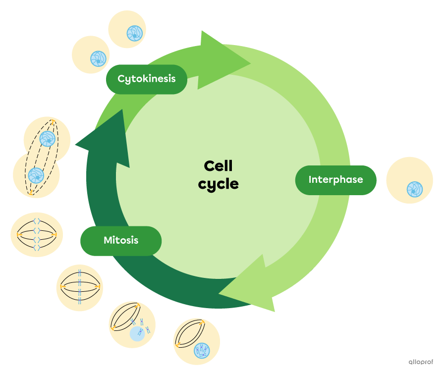
The cell cycle begins with the birth of a cell and concludes at the end of its division. A cell undergoes several changes that are grouped into 3 main stages.
Here are the different forms that the DNA of a human diploid cell assumes during the cell cycle.
| DNA form | Description | Cell cycle stages or phases of mitosis |
|---|---|---|
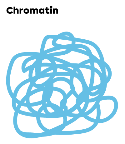 |
In combination with proteins, the DNA is found in the form of threads inside the nucleus. DNA is in the form of chromatin when the cell is not in mitosis. There are usually 46 chromatin threads. |
|
 |
DNA is in the form of threads in the nucleus, but its quantity is doubled because the cell prepares for mitosis. It is the replicated chromatin. The 46 chromatin threads replicate to result in 92 threads. |
|
 |
The replicated chromatin threads coil and condense to form chromosomes composed of 2 sister chromatids. They are genetically identical. The 92 chromatin threads are organized into 46 chromosomes (46 pairs of sister chromatids). Sister chromatids are bound by 2 kinetochores at a centromere. This link is what gives the chromosome its X shape. This shape is clearly visible under a light microscope during the first phases of mitosis. |
|
 |
Individual chromatids are visible under the microscope during the later stages of mitosis. The 46 pairs of sister chromatids separate to form 92 individual chromatids. These 92 chromosomes will be split between 2 nuclei containing 46 chromosomes each, which is the genetic makeup of a diploid cell. |
|
When reading the information in the previous table, it is important to pay attention to the difference between chromatin and chromatid.
Interphase is the longest phase of the cell cycle and it occurs before mitosis.
The interphase itself is divided into 3 phases: G1, S and G2.
| Interphase phase | Description |
|---|---|
| G1 | The G1 phase is a period of growth. The cell maintains its structures, produces new organelles and performs its specific metabolic functions. At this stage, the cell DNA is organized into chromatin. It is also during the G1 phase that the duplication of centrioles is initiated in the cytoplasm. |
| S | The S phase is the period when each chromatin filament in the nucleus replicates. Centrioles continue their duplication process in the cytoplasm. The cell continues to grow. |
| G2 | The G2 phase is a short period of growth. The production of organelles continues, and the duplication of centrioles ends. This is the last phase before the cell enters mitosis. |
Mitosis is a cell division process that allows the division of the cell nucleus into 2 genetically identical diploid nuclei.
Mitosis follows interphase and precedes cytokinesis in the cell cycle.
Mitosis is divided into 5 phases that follow each other continuously without interruption.
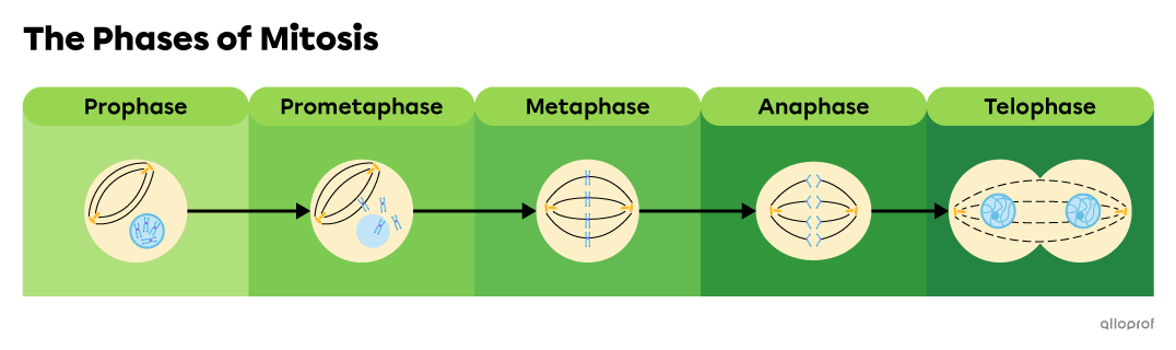
Note: Prometaphase is often included within prophase in textbooks.
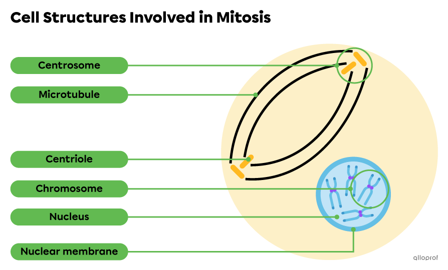
The nucleus, nuclear membrane, centrosomes, centrioles and microtubules are the main cell structures involved in mitosis.
| Structure | Description |
|---|---|
| Nucleus | The nucleus contains the DNA of the cell, among other things. It is surrounded by the nuclear membrane. It is in the nucleus that DNA replication occurs during the S phase of interphase. It is also within the nucleus that the replicated chromatin condenses into chromosomes (sister chromatids) during prophase. |
| Nuclear membrane | The nuclear membrane is a membrane separating the contents of the nucleus from the cytoplasm of the cell. It breaks down during prometaphase and is rebuilt during telophase. |
| Centrosomes and centrioles | The centrosome is an organelle that organizes microtubules during metaphase and facilitates the transport of chromatids during anaphase. A centrosome consists of a pair of centrioles. The centrosome duplicates during interphase (2 centrosomes = 4 centrioles). The centrioles are shaped like small rods arranged perpendicularly to each other. |
| Microtubules | Microtubules are hollow filaments that form in the cytoplasm and organize into spindle fibres. Their role is to align chromosomes (sister chromatids) in the centre of the cell during metaphase and to pull individual chromatids to each pole of the cell during anaphase. |
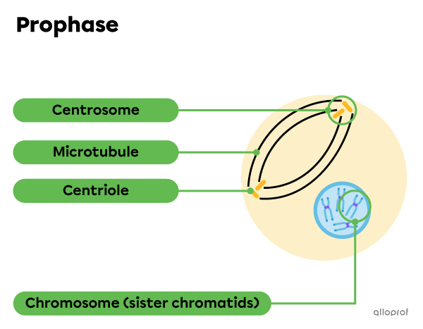
Prophase is the first phase of mitosis.
Here are the main changes that the cell undergoes during prophase.
Threads of replicated chromatin condense to form pairs of sister chromatids. A pair of sister chromatids joined at their centre forms an X shape. This compact organization of the DNA prevents it from tangling and disrupting nucleus division.
Centrosomes and their centrioles move towards the opposite poles of the cell.
Microtubules begin to form near the centrioles.
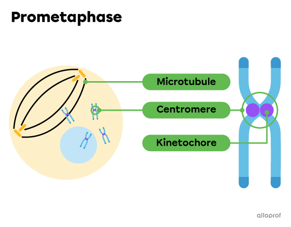
Prometaphase is often included within prophase in most textbooks.
Here are the main changes that the cell undergoes during prometaphase.
The nuclear membrane breaks down and the chromosomes are released into the cytoplasm of the cell.
Kinetochores begin to interact with microtubules.
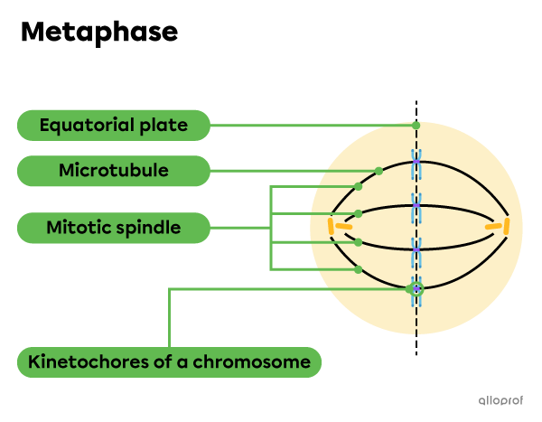
Metaphase is the phase following prometaphase and preceding anaphase.
During metaphase, chromosomes line up in the middle of the cell.
Here are the main changes that the cell undergoes during metaphase.
Kinetochores are attached to microtubules.
Chromosomes migrate to the centre and align along the metaphase plate, sometimes called the equatorial plate.
Microtubules are organized in an elongated spindle apparatus structure, called the mitotic spindle.
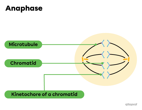
Here are the main changes that the cell undergoes during anaphase.
The kinetochores of sister chromatids separate.
Individual chromatids are carried along microtubules to the opposite poles of the cell.
Cytokinesis begins.
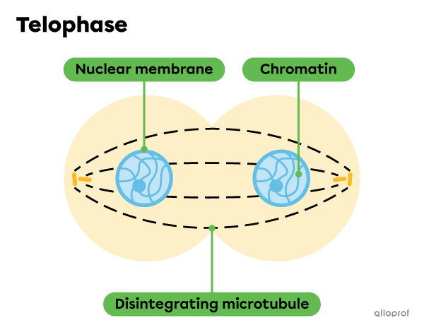
Telophase is the last phase of mitosis.
During telophase, the division of the nucleus is terminated.
Here are the main changes that the cell undergoes during telophase.
Chromatids unfold into chromatin.
A new nuclear membrane is formed around each nucleus.
Microtubules disintegrate.
Cytokinesis continues until completion.
Cytokinesis completes cell division by separating the cytoplasm of the original mother cell and obtaining 2 genetically identical daughter cells. Cytokinesis is initiated during anaphase and continues until completion during telophase.
Finally, each of the new daughter cells enters interphase and a new cell cycle begins.