Subjects
Grades
Meiosis is a cell division process that enables the formation of gametes.
The content covered in this concept sheet goes beyond high school material. It is an enrichment for those who are curious to learn more in depth details. For the content covered in Secondary 3, take a look at this concept sheet.
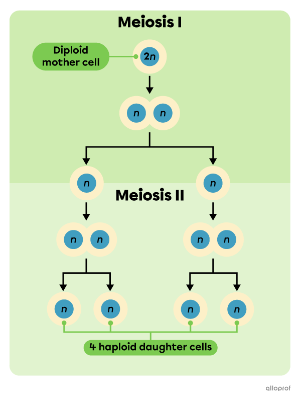
Meiosis is the process that follows the interphase in the cell cycle. Meiosis consists of two consecutive rounds of division: meiosis I and meiosis II. Both processes of nuclear division are followed by cytokinesis, which is the separation of the cytoplasm. Therefore, meiosis I produces two daughter cells from which meiosis II produces four daughter cells.
Meiosis is a reductive cell division, meaning that the genetic material of the daughter cells is reduced compared to that of the mother cell. In fact, the mother cell |\text{2n}| (diploid) produces four daughter cells |\text{n}| (haploid). The four daughter cells obtained by meiosis are genetically different from the mother cell and from each other. The genetic distinction of the four daughter cells promotes genetic diversity of the species through sexual reproduction.
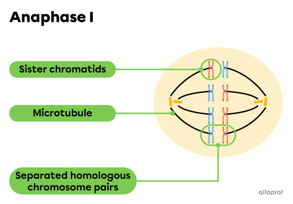
During anaphase I, homologous chromosomes are separated.
During anaphase II, sister chromatids are separated.
A cell undergoing meiosis goes through multiple changes, mainly in its genetic material (DNA). Chromatin, replicated chromatin, sister chromatids and the tetrad of homologous chromosomes are the different forms that the DNA of a cell assumes before and during meiosis.
The genetic material in every cell in the human body is inherited from its parents. In this concept sheet, the genetic material shown in orange comes from Parent 1 and the genetic material shown in blue comes from Parent 2.
Chromatin is the DNA form that resembles threads. It is present when the nucleus is not yet in the process of cell division.
Chromatin threads
Replicated chromatin is found in the cell during the interphase. During this phase, the DNA still appears as threads inside the nucleus, but its amount has doubled, because the cell is preparing for meiosis I.
Threads of replicated chromatin
The threads of replicated chromatin coil and condense to form chromosomes composed of 2 genetically identical sister chromatids.
The sister chromatids are linked at the centromere by their kinetochore, which is a structure that binds microtubules during prophase. This link is what gives the chromosome its X shape.
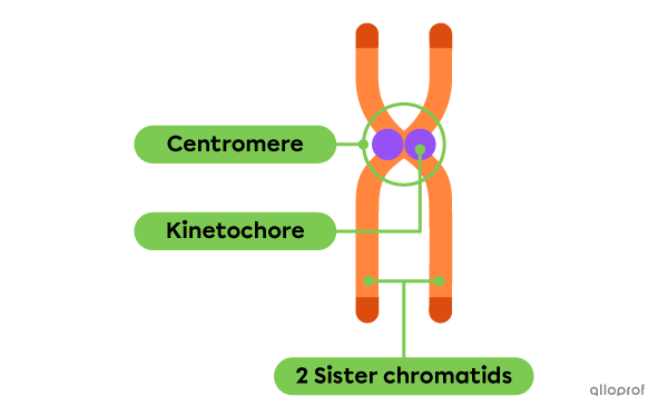
A pair of sister chromatids from Parent 1
Homologous chromosomes are pairs of sister chromatids containing the genetic material that codes for the same genes. Homologous chromosomes are not genetically identical to each other since one pair comes from Parent 1 and the other pair comes from Parent 2.
The two homologous chromosomes grouped together are called a tetrad, where tetra- means four. A tetrad consists of one pair of homologous chromosomes for a total of four chromatids. In other words, a tetrad consists of two pairs of sister chromatids for a total of four chromatids.
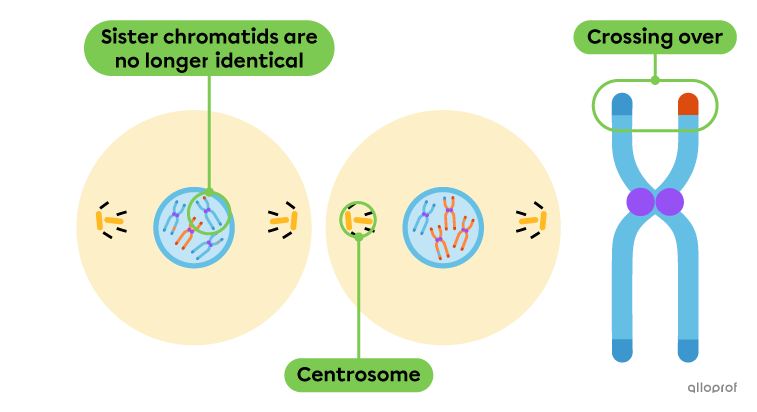
The non-sister chromatids in the centre of the tetrad tend to exchange genetic material. This phenomenon is known as chromosomal crossover, or crossing over. Once crossing over occurs, the sister chromatids are no longer identical to each other.
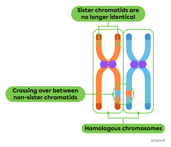
A tetrad of homologous chromosomes
Crossing over is an exchange of genes or alleles between the two non-sister chromatids at the centre of a tetrad. The process of crossing over takes place during prophase I.
When two homologous chromosomes come together in a tetrad, their chromatids align and come into contact with each other. Through this contact, genes are swapped between the non-sister chromatids in the middle of the tetrad.
As a result of crossing over, each tetrad generates four chromatids that are genetically different from each other. This difference contributes to increasing the genetic diversity of the species.
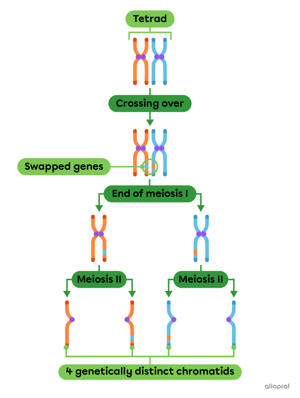
The effect of crossing over on the chromatids obtained by the end of meiosis
The nucleus, nuclear membrane, centrosomes, centrioles and microtubules are the main cell structures involved in meiosis.

| Structures | Description |
|---|---|
| Nucleus | The nucleus contains the cell's DNA, among other things. It is surrounded by the nuclear membrane. Replication of DNA prior to meiosis I occurs inside the nucleus. |
| Nuclear membrane | The nuclear membrane is a barrier that separates the contents of the nucleus from the cytoplasm of the cell. |
| Centrosomes and centrioles | The centrosome is an organelle that organizes microtubules and facilitates the transport of chromosomes. A centrosome consists of a pair of centrioles. A dividing cell has 2 centrosomes for a total of 4 centrioles. The centrioles are shaped like small rods arranged perpendicularly to each other. |
| Microtubules | Microtubules are hollow filaments that form in the cytoplasm and are organized into spindle fibres. Their role is to align the chromosomes in the centre of the cell and pull them to each pole of the cell. |
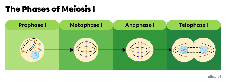
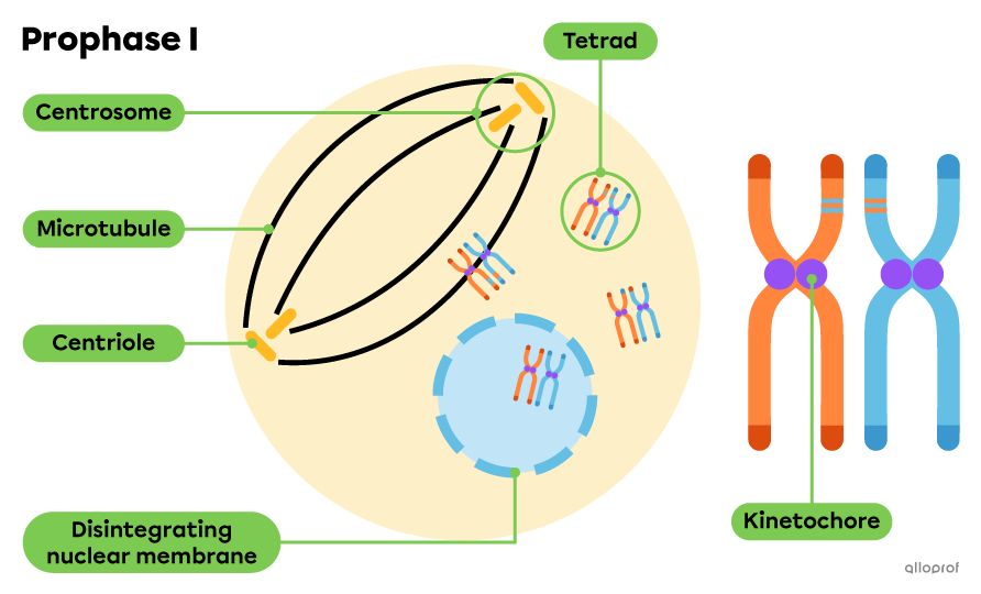
Here are the main changes in the cell during prophase I:
Replicated chromatin threads condense inside the nucleus to form pairs of sister chromatids. They join their homologous pair to form tetrads.
Crossing over occurs.
The nuclear membrane is broken down and the tetrads are released into the cytoplasm of the cell.
Centrosomes and their centrioles move to opposite poles of the cell.
Microtubules begin to form near the centrioles.
Kinetochores begin to engage with the microtubules.
Here are the main changes that the cell undergoes during metaphase I:
Kinetochores attach to microtubules.
Microtubules organize themselves into an elongated spindle apparatus, called a meiotic spindle.
The tetrads of homologous chromosomes align randomly along the metaphase plate, sometimes called the equatorial plate, in the centre of the cell.
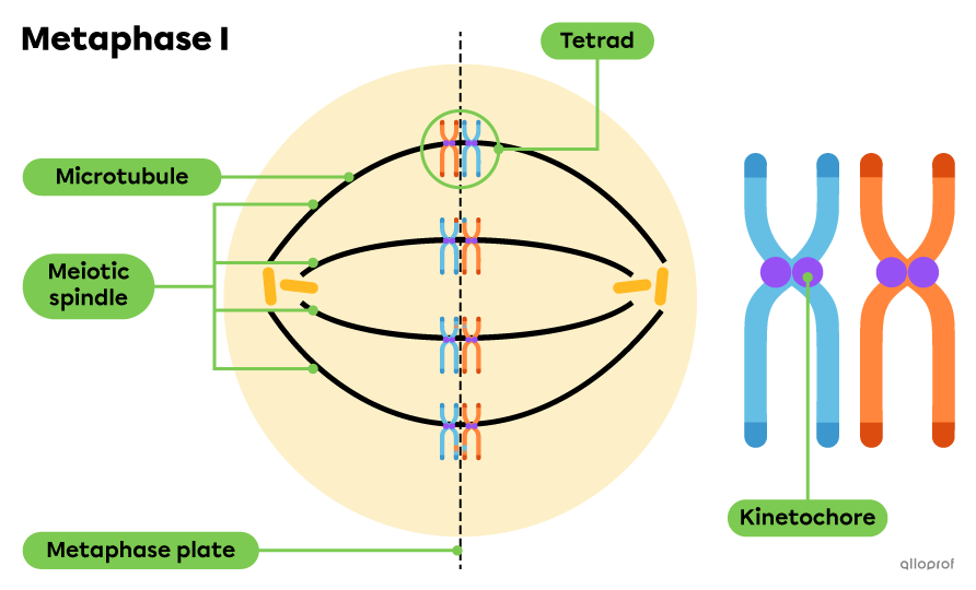
Note: In this picture, the sister chromatids of Parent 1 and Parent 2 are randomly aligned. More genetic material from Parent 1 goes to the right pole of the cell and more genetic material from Parent 2 goes to the left pole of the cell. This random alignment determines the genetic background of the two cells obtained by the end of meiosis I.
Here are the main changes that the cell undergoes during anaphase I:
The homologous chromosomes separate, but the sister chromatids remain linked by their kinetochore.
The sister chromatids are pulled along the microtubules towards each pole of the cell.
The cell is slightly elongated, indicating that the first round of cytokinesis is beginning.
Here are the main changes in the cell during telophase I:
The nuclear membrane temporarily reforms around the two new nuclei.
The microtubules disintegrate.
The first round of cytokinesis continues and is completed shortly after telophase I.
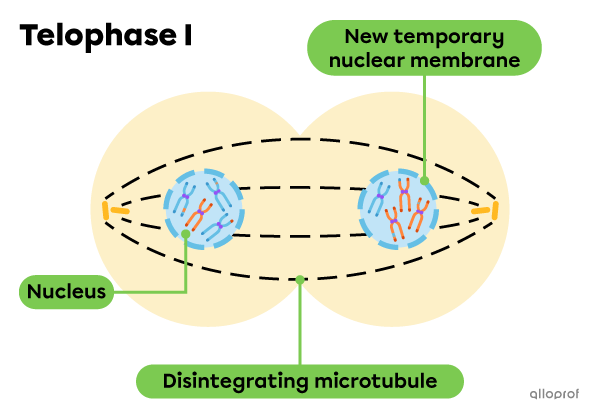
When the first round of cytokinesis is completed, two haploid daughter cells |(\text{n}),| whose DNA is organized into pairs of sister chromatids, are obtained.
However, due to crossing over, the sister chromatids are usually no longer identical to each other.
Two daughter cells obtained by the end of meiosis I and cytokinesis.
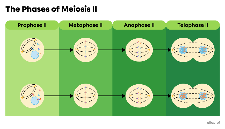
Between the end of meiosis I and the beginning of meiosis II, there is no replication of genetic material.
The centrosomes present in the two daughter cells will duplicate before the start of meiosis II because two centrosomes per cell are required to initiate it.
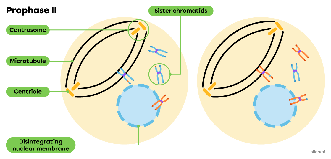
Here are the main changes that each daughter cell undergoes during prophase II:
The temporary nuclear membrane breaks down to release sister chromatids into the cytoplasm.
Centrosomes and their centrioles move to opposite poles of the cell.
The kinetochores of the sister chromatids begin to engage with the microtubules.
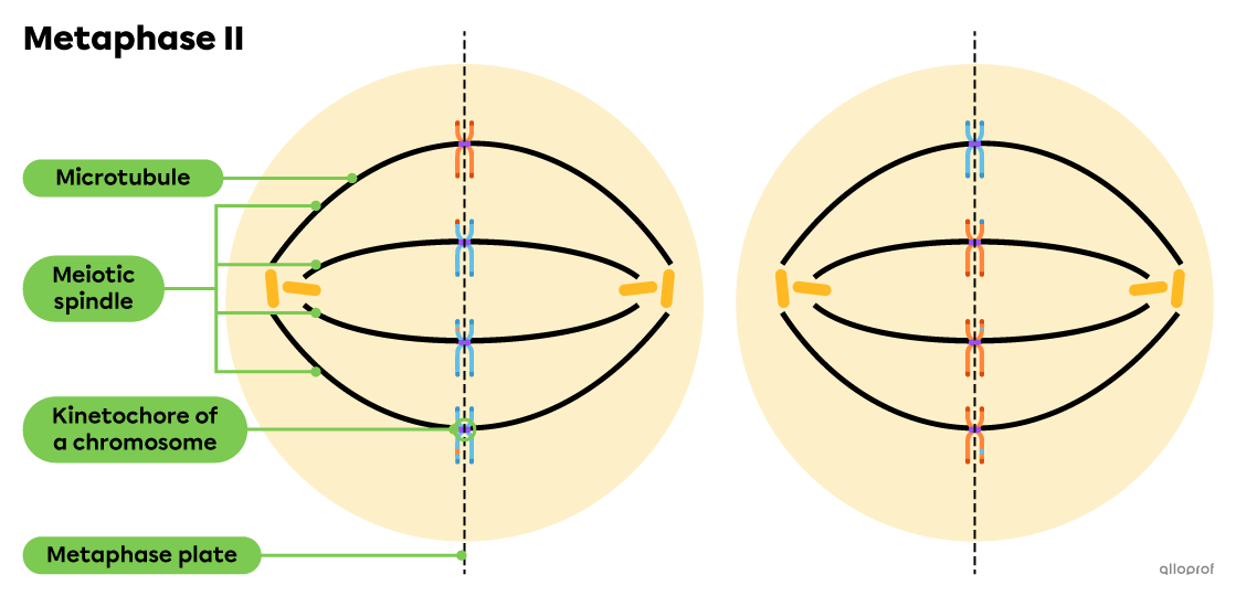
Here are the main changes that each daughter cell undergoes during metaphase II:
Kinetochores attach to microtubules.
The microtubules organize themselves into an elongated spindle apparatus, called a meiotic spindle.
Sister chromatids align randomly on the metaphase plate, sometimes called the equatorial plate.
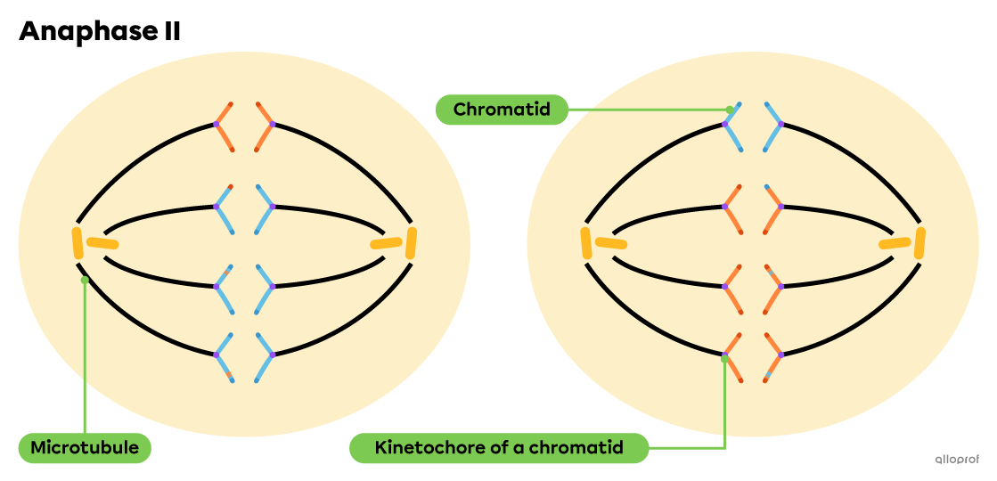
Here are the main changes that each daughter cell undergoes during anaphase II:
The kinetochores of the sister chromatids separate.
The chromatids are pulled along the microtubules towards each pole of the cell.
The cell is slightly elongated, indicating that the second and final round of cytokinesis is beginning.
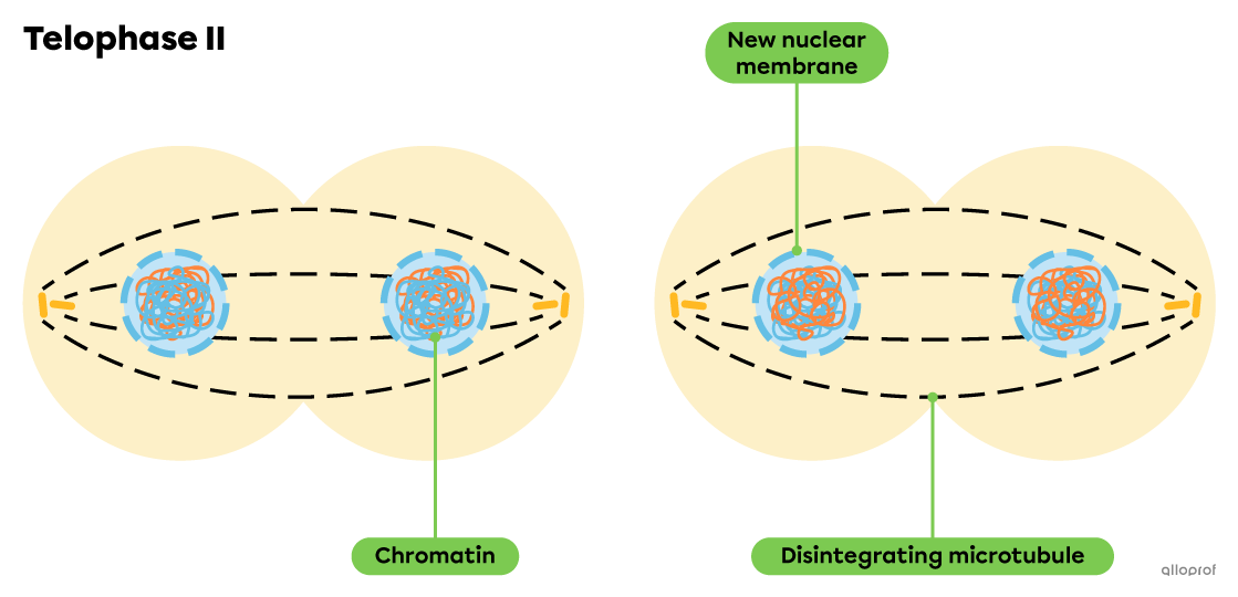
Here are the main changes that each daughter cell undergoes during telophase II:
Chromatids unwind into chromatin.
New nuclear membranes form around two new nuclei.
The microtubules disintegrate.
The second round of cytokinesis continues and is completed shortly after telophase II.
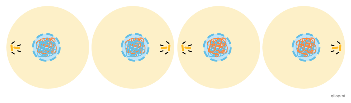
Four daughter cells obtained after meiosis II and cytokinesis